Clostridioides Difficile Colitis
Clostridioides Difficile Colitis
- Bacteriology
- C. difficile is a gram-positive, spore-forming, toxin-producing, anaerobic bacteria
- most commonly associated with antibiotic use and disruption of the normal microbiome
- pathogenetic strains produce two toxins (A and B) that are responsible for the symptoms of
C. difficile infection (CDI)
- hypervirulent strains have emerged that produce 16X to 20X more toxin than less virulent strains
- pathogen most responsible for nosocomial infectious diarrhea
- Infection Risk Factors
- prior treatment with antibiotics, especially fluoroquinolones, clindamycin, cephalosporins, and penicillins
- antibiotics suppress the normal colon flora, allowing overgrowth of C. difficile
- other risk factors include age > 65, current or recent hospitalization, and use of proton pump inhibitors
- Clinical Presentation
- infection (CDI) can vary from mild diarrhea to fulminant infection with toxic megacolon and systemic sepsis
- infection typically begins within 4 to 9 days after starting antibiotics
- 25% of patients may present up to 10 weeks after a course of antibiotics
- Mild Disease
- main symptom is watery diarrhea – more than 3 loose stools / 24 hrs
- other symptoms include lower abdominal pain and cramping, low-grade fever, anorexia, and nausea
- physical exam may demonstrate lower abdominal tenderness
- average WBC count is 15,000
- Severe Disease
- diffuse abdominal pain and distention
- hypovolemia, elevated creatinine, and lactic acidosis are common
- WBC count may be as high as 40,000
- Fulminant Disease
- hypotension may progress to shock and multisystem organ failure
- toxic megacolon
- bowel perforation with peritonitis
- WBC count may be > 50,000
- Recurrent Disease
- 25% of patients relapse within 30 days of completing treatment
- recurrent disease can be mild, severe, or fulminant
- one major risk factor for recurrence is the ongoing need for concomitant antibiotics
during treatment for CDI
- Asymptomatic Carriers
- 20% of hospitalized patients are asymptomatic carriers
- no need to screen for these patients, and no need for treatment or contact precautions
- Diagnosis
- Stool Studies
- CDI is diagnosed by a positive test for C. difficile toxins or the C. difficile toxin B gene
- there are several diagnostic tests available, each with their advantages and disadvantages
- GDH Antigen Test
- uses antibodies to test for the GDH enzyme, a protein present in all C. difficile species
- cheap and fast
- a negative test is useful (good sensitivity)
- a positive test, however, cannot distinguish between toxigenic and nontoxigenic strains
(poor specificity)
- Toxin A and B Test
- low cost, fast turnaround
- both toxins should be tested for
- a positive test is valuable (high specificity)
- high rate of false negatives (low sensitivity)
- Interpretation of Results
- if both tests are positive, then the patient should be treated for CDI
- if both tests are negative, then the patient does not have CDI
- discordant results (one test positive, the other negative) should be resolved with the NAAT test
- NAAT Test
- polymerase chain reaction test for the gene encoding toxin B
- a positive test is considered evidence of CDI, as long as one of the first two tests was also positive
- Endoscopy
- not usually necessary unless another diagnosis is suspected that requires direct visualization
and/or biopsy
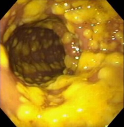
- flexible sigmoidoscopy or colonoscopy may identify the plaque-like pseudomembranes which are pathognomonic
for CDI
- pseudomembranes are found in 25% of patients with mild disease, and 87% with fulminant colitis
 Pseudomembranes
- Management
- Medical Management
- Infection Control
- initiate hospital infection control practices
- contact precautions
- hand-washing with soap and water may be more effective than alcohol-based hand sanitizers since
C. difficile spores are resistant to alcohol
- Antibiotic Management
- discontinue the offending antibiotics if possible
- if continued antibiotic treatment is necessary, then switching to other agents less frequently
implicated in CDI is prudent
- Diarrhea Management
- correct fluid and electrolyte losses
- antimotility agents (loperamide and opiates) are usually avoided, but the evidence that they
cause harm is not strong
- patients can have a regular diet as tolerated
- Treatment
- Mild Disease
- oral vancomycin is the preferred antibiotic
- oral fidaxomicin and oral metronidazole are alternatives
- treatment course is for 10 days
- Severe or Fulminant Disease
- higher doses of antibiotics are used
- a second agent may be added if no response
- drugs may need to be given via an NG tube
- in refractory cases, vancomycin can be given as an enema, but is associated
with perforation
- Surgery
- Indications
- toxic megacolon
- perforation
- failure of medical management with ongoing systemic toxicity
- Procedures
- Total Abdominal Colectomy with End ileostomy
- indicated for perforation, necrosis, or abdominal compartment syndrome
- residual infection in the rectal pouch may be managed with vancomycin flushes
through a rectal tube
- since CDI is a pancolonic disease, segmental colectomy is not indicated for isolated
areas of perforation or necrosis
- Diverting Loop Ileostomy with Colonic Lavage
- since the mortality rate of abdominal colectomy and ileostomy is so high (34% to 57%),
other surgical options have been explored
- one promising procedure is laparoscopic loop ileostomy creation with intraoperative
antegrade colonic lavage using polyethylene glycol
- the fluid is collected with a rectal tube
- postoperatively, vancomycin irrigation through the ileostomy is continued for 10 days
- associated with lower mortality and higher ileostomy reversal rates in nonrandomized
studies than total abdominal colectomy
Ischemic Colitis
- Anatomy
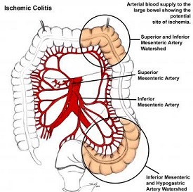
- colon receives its blood supply from the SMA and IMA
- SMA gives off the ileocolic, right colic, and middle colic arteries
- IMA gives off the left colic, sigmoid, and superior rectal arteries
- rectum also gets blood supply from the iliac arteries via the inferior and middle rectal vessels
- SMA and IMA have an extensive collateral network that limits the risk of colon ischemia
- Collateral Connections
- marginal artery of Drummond forms a continuous arcade near the colon wall
- in some patients, the arc of Riolan connects the SMA, via the middle colic, with the IMA, via the left colic
- Watershed Areas
- regions of the colon that have limited collateral blood flow
- splenic flexure (Griffith's point) is where the SMA and IMA circulations meet
- rectosigmoid junction (Sudek's point) is where the distal sigmoid arteries and superior rectal artery meet

- Etiologies
- Occlusive Causes
- thromboembolism, usually from atrial fibrillation
- IMA ligation during aortic reconstruction
- Nonocclusive Causes
- accounts for 95% of cases of colon ischemia:
- heart failure
- arrhythmias
- shock
- vasopressors
- cardiopulmonary bypass
- Classification
- Partial Thickness
- ischemia limited to the mucosa and submucosa
- usually resolves with nonoperative management
- may result in stricturing and scarring
- Full Thickness
- perforation is common
- urgent surgery is necessary
- Clinical Presentation
- left-sided abdominal pain, cramping, low-grade fever, bloody diarrhea is typical of partial thickness disease
- full thickness ischemia can result in peritonitis, high fever, leukocytosis, acidosis
- Diagnosis
- Plain Films
- most valuable for detecting free air or pneumatosis, and for ruling out obstruction
- Lab Studies
- no specific marker for ischemia
- elevated lactate, LDH, CPK, or amylase suggest significant tissue damage
- CT Scan
- best study is with IV and oral contrast, if possible
- valuable for ruling out nonischemic causes of abdominal pain
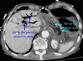
- suggestive, but nonspecific, findings of ischemia include colon wall thickening,
pneumatosis, pericolonic fat stranding, portal venous air

- Endoscopy
- allows direct vision of the colon mucosa
- cannot reliably distinguish between mucosal ischemia and full-thickness ischemia
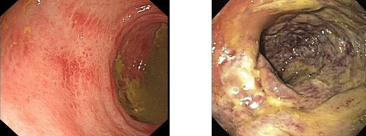
- partial thickness ischemia is suggested by hemorrhagic, pale mucosa with patches
of inflammation interspersed between areas of healthy mucosa
- full thickness ischemia is suggested by dusky mucosa, submucosal hemorrhage,
hemorrhagic ulcerations or frank necrosis
 (L) Partial thickness ischemia
(R) Full thickness ischemia
- Angiography
- only role is if embolism is suspected
- Treatment
- Nonoperative Management
- nearly 80% of patients respond to conservative therapy:
- NPO
- fluid resuscitation for low flow states
- NG tube for nausea, vomiting
- broad-spectrum antibiotics to prevent translocation of bacteria
- discontinue vasopressors if possible
- goal is to maximize cardiac output and mesenteric blood flow
- failure to improve within 24 – 48 hours should mandate repeat CT scan or endoscopy
- Surgery
- indications include peritonitis, free air, massive hemorrhage, or evidence of full thickness
necrosis on imaging or endoscopy
- revascularization procedures are not indicated
- open or laparoscopic segmental resection with an ostomy or mucous fistula is the usual procedure
- primary anastomosis should be avoided in this patient population
- consider a second look procedure if there is concern for ongoing ischemia
- if the entire colon is involved, then total colectomy and ileostomy is necessary
References
- Sabiston, 20th ed., pgs 1353 – 1359
- Cameron 11th ed., pgs 169 – 172, 173 – 177
- UpToDate. Clostridioides (formerly Clostridium) Difficile Infection in Adults: Treatment and Prevention.
Ciaran P. Kelley MD, et. al. Jan 30, 2020. Pgs 1 – 35.
- UpToDate. Surgical Management of Clostridioides (Formerly Clostridium) Difficile Colitis in Adults.
Brian Zuckerbraun, MD. Jan 21, 2019. Pgs 1 - 16.
- UpToDate. Colonic Ischemia. Peter Grubel, MD, et. al., Mar 05, 2020. Pgs 1 – 35.


