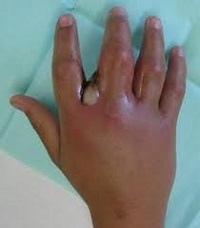
Hand infections


Superficial Hand Infections

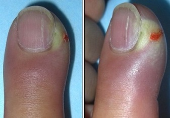
  1. Paronychia
    1. Etiology
      • infection of the nail bed or periungual soft tissues
      • trauma, manicures, nail biting are common causes
      • infection usually begins on the side of the nail bed and can spread beneath the nail if left untreated
      • most common organism is S. aureus

    2. Clinical Presentation
      • extremely painful if pus is trapped beneath the nail

      Paronychia
    3. Management
      • initial treatment is antibiotics and warm compresses
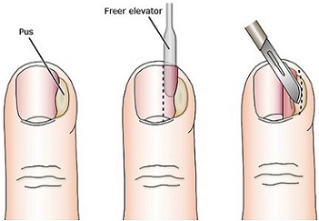
      • if an abscess develops, then drainage is required
      • if the abscess is lateral to the nail, then an incision is made over the fluctuance
      • if the nail bed is involved, then partial or complete removal of the nail bed is required

      Drainage of a Paronychia
  2. Pulp Space Infections (Felons)
    1. Etiology
      • closed space infection usually caused by a puncture wound or splinter
      • thumb and index finger are most commonly affected

    2. Clinical Presentation
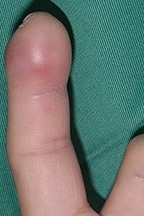
      • rapid onset of throbbing pain in the affected pulp space
      • swelling and exquisite tenderness are also present

      Felon - Finger
    3. Management
      • surgical emergency
      • tension in the pulp space can result in gangrene of the fingertip and necrosis of the terminal phalanx
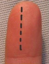
      • longitudinal incision is made over the area of fluctuance
      • to avoid a flexion contracture, the incision shouldn’t cross the distal interphalangeal joint
      • wound is loosely packed and a finger splint applied

      Felon Drainage
  3. Web Space Abscesses
    1. Etiology
      • direct implantation or spread from a pulp space infection

    2. Clinical Presentation
      • fluctuant mass in the web space separates the fingers
      • there is loss of the normal palmar concavity with a widened space between the fingers

      Webspace Abscess
    3. Management
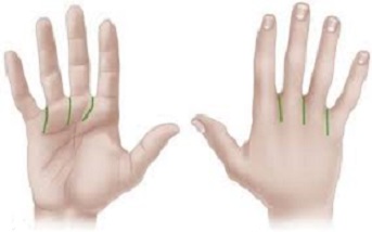
      • may require both a volar incision and a dorsal incision

      Webspace Abscess Drainage
  4. Herpetic Whitlow
    1. Etiology
      • viral infection caused by type 1 or type 2 herpes simplex virus
      • occupational hazard in healthcare workers

    2. Clinical Presentation
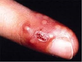
      • infection begins with small clear vesicles
      • a single finger is usually painful, erythematous, and swollen
      • vesicles coalesce by day 14 and ulcerate
      • may be mistaken for a paronychia

      Herpetic Whitlow
    3. Management
      • surgical drainage is contraindicated because it can lead to spread of the virus
      • treatment is supportive
      • antivirals may be of help if they are started within 48 hours of symptoms

Deep Space Infections

  1. Anatomy
    • deep space infections involve the tendons and structures deep to the tendon sheaths
    • usually occur as a result of direct implantation
    • in the early stage, the infection is limited by the compartments of the hand
    • as the infection progresses, it may rupture superficially, spread to adjoining compartments, or involve the underlying bone and joints
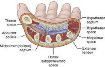
    Deep Space Infections Anatomy
  2. Palmar Space Infections
    1. Thenar Abscesses
      1. Clinical Presentation
        • characterized by a widely abducted thumb and fullness on the dorsum of the first web space
        • severe pain on adduction or opposition of the thumb
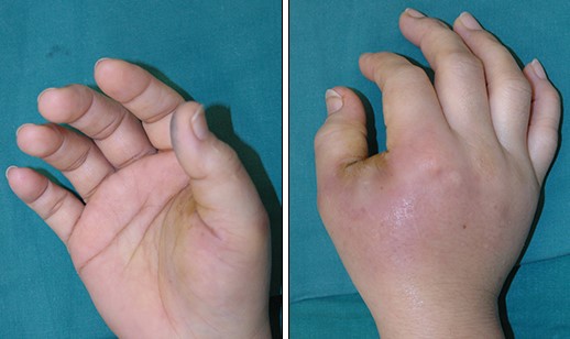
        Thenar Space Abscess
      2. Management
        • requires both a volar and dorsal incision
        • the volar incision is parallel to the thenar crease
        • palmar cutaneous branch of the median nerve is at risk in the proximal part of the volar incision
        • the motor branch of the median nerve is also at risk
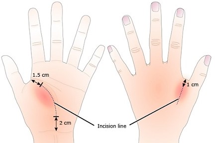
        Thenar Space Abscess Drainage
    2. Midpalmar Space Abscesses
      1. Clinical Presentation
        • loss of the normal palmar concavity
        • the middle and ring fingers assume a partially flexed posture
        • pain on passive extension of these fingers
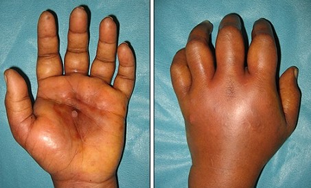
        Midpalmar Space Abscess
      2. Management
        • drainage is accomplished with a longitudinal or curvilinear incision
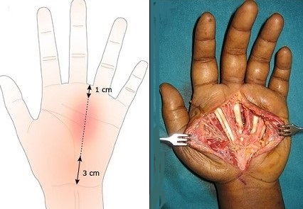
        Midpalmar Space Abscess Drainage
    3. Hypothenar Space Abscesses
      1. Clinical Presentation
        • no involvement of the fingers or flexor tendons
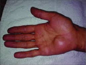
        Hypothenar Space Abscess
      2. Management
        • longitudinal incision over the area of greatest fluctuance
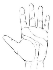
        Hypothenar Space Abscess Drainage
  3. Flexor Tenosynovitis
    1. Etiology
      • the flexor tendons of the hand are enclosed in a double layered synovial sheath
      • the synovial spaces intercommunicate, are poorly vascularized, and are rich in synovial fluid, allowing for an optimal environment for bacterial growth
      • bacterial proliferation leads to increased pressure within the sheath, which obstructs blood flow and can cause tendon necrosis and rupture
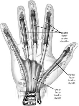
      Flexor Tendon Sheaths
    2. Clinical Presentation
      1. Kanavel’s Four Cardinal Signs
        • affected finger is partially flexed
        • symmetrical swelling of the entire finger
        • tenderness along the flexor tendon sheath
        • exquisite pain on passive extension of the finger
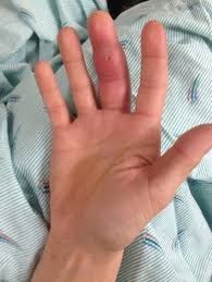
        Hypothenar Space Abscess Drainage
    3. Management
      1. Nonoperative Management
        • appropriate for patients seen within 24 hours of infection
        • consists of antibiotics, arm elevation, hand immobilization

      2. Operative Management
        • indicated if nonoperative management is unsuccessful or if the infection is greater than 48 hours old
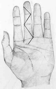
        Drainage of Flexor Tenosynovitis


References

  1. Sabiston, 20thed., pgs 1999 – 2002
  2. UpToDate. Overview of Hand Infections. Sandeep J. Sebastin Muttah, MMed, FAMS, Kevin C. Chung, MD, MS, Shimpei Ono, MD, PhD. Jan 02, 2020. Pgs 1 - 69
  3. SESAP. 17th ed. Soft Tissues