Basal Cell Carcinoma (BCCA)
- Epidemiology
- most common skin cancer
- exposure to UV radiation is the most common risk factor
- 90% have a mutation in the hedgehog signaling pathway
- significant occupational hazard for people with outdoor occupations
- more common in people with fair complexions
- Clinical Appearance
- several different morphologies:
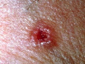
- Nodular BCCA
- most common type (70%)
- waxy, cream-colored lesion with rolled borders
- often contains a central ulcer (“rodent ulcer”)
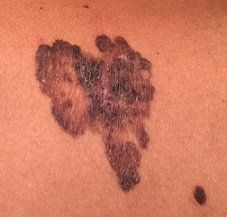
- Pigmented BCCA
- tan to black in color
- must be distinguished by biopsy from melanoma
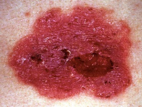
- Superficial BCCA
- red, scaly lesion with irregular, ill-defined margins
- may be mistaken for psoriasis or eczema
- usually found on the trunk
- Clinical Course
- usually slow growing
- often neglected for years
- metastasis and death are extremely rare
- can cause extensive local destruction
- Risk of Recurrence
- BCCAs can be classifies as low risk or high risk for recurrence
- Low Risk
- lesions < 10 mm on the cheek, forehead, scalp, neck
- lesions < 20 mm on the trunk and extremities
- well-defined borders
- no history of immunosuppression or radiation treatment at the site
- High Risk
- lesions >= 10 mm on the face or >= 20 mm on the trunk and extremities
- recurrent lesions
- history of immunosuppression or radiation
- perineural or vascular invasion
- Treatment
- Surgical Excision
- 4 – 6 mm margins are adequate for low-risk lesions
- high-risk lesions require 10 mm margins
- primary closure is usually possible
- Mohs Micrographic Surgery
- used for low-risk lesions on cosmetically sensitive areas of the face (eyelids, nose)
- involves serially excising the lesion with immediate frozen-section examination of all the margins
- major drawbacks are cost and the length of time involved (up to several days), since complete excision
may require multiple attempts
- cure rates are comparable to wide excision
- Topical Treatments
- usually offered to patients who prefer to avoid excision
- imiquimod or topical 5-fluoruracil are associated with higher recurrence rates than surgery
- Field Therapy
- treats a generalized area
- margins cannot be evaluated
- many options are available: cryotherapy, radiation therapy, electrodessication and curettage,
and photodynamic therapy
Squamous Cell Carcinoma (SCCA)
- Epidemiology
- second most common skin cancer
- risk factors include sun exposure, chemical carcinogens, previous radiation, chronic inflammation
(burn scars, chronic osteomyelitis) and chronic immunosuppression
- after organ transplantation, there is a 65 times increased risk of developing a SCCA
- invasive SCCAs may metastasize to regional lymph nodes or distant sites
- Biologic Behavior
- may spread both vertically and horizontally
- Tumor Thickness
- correlates well with biologic behavior
- lesions that recur locally are usually more than 4 mm thick
- lesions that metastasize are usually more than 10 mm thick
- Tumor Size
- tumors less than 2 cm in diameter have recurrence rates of 5% and a 7% incidence of metastasis
- tumors greater than 2 cm have twice the local recurrence rate and three times the incidence of
regional lymphatic spread
- Tumor Location
- tumors arising in burn scars, areas of chronic osteomyelitis, and areas of previous injury metastasize early
- lesions on the external ear frequently recur and involve regional nodes early
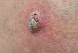
- Clinical Appearance
- scaly lesions that become ulcerated centrally and have elevated, firm edges
- may be confused with keratoacanthoma
- Treatment
- Primary Lesion
- small lesions may be treated with curettage/electrodessication, cryotherapy, or topical agents
- most low-risk lesions should be surgically excised with a 4 – 6 mm margin
- high-risk lesions will require a 1 cm margin
- Mohs surgery is indicated for lesions that require a tissue-sparing approach (eyelid, nose)
- Regional Nodes
- a SLN biopsy is indicated in high-risk patients with clinically negative nodes
- a node dissection is indicated for clinically palpable nodes
- a prophylactic node dissection is indicated for lesions arising in chronic wounds
Merkel Cell Carcinoma (MCC)
- Epidemiology
- rare, aggressive cutaneous malignancy with a high risk of recurrence and metastasis
- primary affects older adults with fair skin
- has a predilection for sun-exposed areas
- more frequent in immunosuppressed patients
- Pathogenesis
- neuroendocrine tumor that may originate from Merkel cells, which are located in the basal layer of the epidermis
and hair follicles
- Merkel cell polyomavirus has been causally linked to the development of MCC in 60% - 80% of patients
- Clinical Manifestations
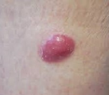
- most present as a fast growing, firm, flesh-colored or reddish-colored, intracutaneous nodule
- majority are located on the head and neck, upper arms, and shoulders
- 26% of patients have regional node involvement, and 8% already have distant metastases at the time of diagnosis
- Management
- Primary Tumor
- wide local excision with 1 - 2 cm margins
- adjuvant XRT, especially for head and neck lesions or close margins
- Regional Lymph Nodes
- Clinically Negative Nodes
- SLN biopsy since occult nodal disease is found in 33% of patients
- completion node dissection and adjuvant XRT is indicated for a positive SLN
- Clinically Positive Nodes
- requires FNA or core needle biopsy confirmation
- complete metastatic work up
- regional node dissection followed by adjuvant XRT if the metastatic workup is negative
- Adjuvant Therapy
- no evidence to support adjuvant chemotherapy or immunotherapy, although these agents have value in
metastatic disease
References
- Sabiston, 20th ed., pgs 747 – 750
- UpToDate. Treatment and Prognosis of Basal Cell Carcinoma at Low Risk of Recurrence. Sumaira Z. Aasi, MD.
July 22, 2020. Pgs 1 – 39
- UpToDate. Treatment and Prognosis of Low-risk Cutaneous Squamous Cell Carcinoma. Sumaira Z. Aasi, MD,
Angela M. Hong, MBBS, MMed, PhD. Oct 28, 2020. Pgs 1 – 41
- UpToDate. Pathogenesis, Clinical Features, and Diagnosis of Merkel Cell (Neuroendocrine) Carcinoma.
Patricia Tai, MB, BS, DABR, FRCR, FRCPC, Paul T Nghiem, MD, PhD, Song Youn Park, MD. Aug 13, 2020.
Pgs 1 – 25
- UpToDate. Staging and Treatment of Merkel Cell Carcinoma. Patricia Tai, MB, BS, DABR, FRCR, FRCPC, Song Youn Park,
MD, Paul T Nghiem, MD, PhD, July 15, 2019. Pgs 1 – 25


