Small Bowel Diverticula
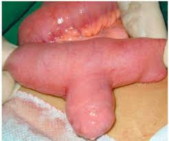
- Meckel’s Diverticulum
- found in 2% of the population
- true diverticulum containing all four bowel wall layers
- usually occurs within 100 cm of the ileocecal valve
- majority contain heterotopic gastric or pancreatic mucosa
- results from failure or incomplete vitelline duct obliteration

- Clinical Manifestations
- asymptomatic unless complications occur
- Bleeding
- most common complication in the pediatric population
- rare complication in older patients
- results from an ileal ulceration adjacent to acid-producing heterotopic gastric mucosa
located within the diverticulum
- Obstruction
- most common complication in adults
- may occur as a result of intussusception with the diverticulum acting as a lead point
- volvulus may occur if there is a fibrous band attaching the diverticulum to the umbilicus
- Diverticulitis
- clinically indistinguishable from appendicitis
- Diagnosis
- in a bleeding pediatric patient, a Meckel’s radionuclide scan is 90% accurate in identifying a
Meckel’s diverticulum
- in symptomatic adults, the majority are identified during laparoscopy or laparotomy
- Surgical Management
- Symptomatic Patients
- bleeding patients should have a segmental resection, including the ileal ulceration
- segmental resection will also be required if the base of the diverticulum is inflamed
or perforated
- diverticulectomy may be possible in patients with obstruction from intussusception
- Incidentally Found Meckel's Diverticulum
- most surgeons do not perform prophylactic diverticulectomy
- however, any bands attaching the diverticulum to the abdominal wall should be lysed to prevent a
future volvulus
- Duodenal Diverticula
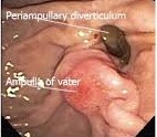
- false diverticula, only containing the mucosa and submucosa
- majority are located near the ampulla on the medial wall of the duodenum and may involve the pancreas
- detected on 5% - 27% of ERCPs

- Pathophysiology
- acquired defect
- results from herniation through the bowel wall at sites where large vessels enter
- dysmotility from increased intraluminal pressures may also play a role
- Clinical Manifestations
- majority are asymptomatic
- rarely, may cause bleeding, biliary obstruction or recurrent pancreatitis
- may cause technical challenges during ERCP for choledocholithiasis
- Management
- asymptomatic diverticula should be left alone
- symptomatic lateral wall diverticula can be managed with diverticulectomy
- symptomatic medial wall diverticula should be managed endoscopically if possible
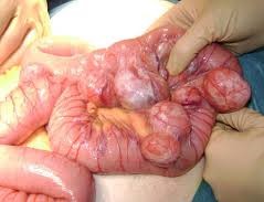
- Jejunal/Ileal Diverticula
- majority are false diverticula
- usually multiple and localized to the proximal jejunum
- often associated with intestinal dysmotility

- Clinical Manifestations
- most are asymptomatic
- some patients develop bacterial overgrowth: bloating, malabsorption, steatorrhea,
vitamin B12 deficiency
- additional complications include bleeding, obstruction, diverticulitis
- Management
- bacterial overgrowth and diverticulitis are treated with antibiotics
- bleeding will require identification of the bleeding site, which may require tagged RBC bleeding scans
and/or angiography
- surgical management of bleeding or obstruction will require segmental resection
Chronic Radiation Enteritis
- Pathophysiology
- Radiation therapy for pelvic cancers may result in a progressive occlusive vasculitis that leads to
chronic ischemia and fibrosis of the small bowel
- These changes can result in strictures, abscesses, and fistulas
- Terminal ileum is the most frequently involved segment
- Clinical Manifestations
- Most patients become symptomatic within 2 years of treatment, although in some patients symptoms do
not develop until many years later
- Partial small bowel obstruction is the most common presentation – crampy abdominal pain, nausea,
and vomiting
- Complete small bowel obstruction, lower GI bleeding, and abscesses and fistulas are less common
presentations
- Diagnosis
- Contrast studies will show luminal narrowing, loss of mucosal folds, and ulceration
- CT scanning is neither sensitive nor specific for radiation enteritis, but it is valuable to rule out
recurrent cancer
- Indications for Surgery
- Obstruction that does not resolve with conservative therapy
- Perforation
- Intra-abdominal abscesses
- Fistulas
- Hemorrhage
- Surgical Management
- Goal is a limited resection with primary anastomosis between healthy bowel segments
- Limited resection, however, is often difficult because of diffuse fibrosis and dense adhesions
- Distinguishing between normal and irradiated bowel can also be challenging at times
- Anastomoses between irradiated segments have a leak rate of 50%
- Mortality rate is 10%
- If a limited resection is not feasible, then an intestinal bypass procedure may be the best option
References
- Schwartz, Principles of Surgery. 10th ed., Pgs 1162 - 1167
- Sabiston, Textbook of surgery. 20th ed., Pgs 1280 - 1286, 1290 - 1291


