Overview
- Incidence
- Rare tumors comprising < 1% of adult malignancies and 12% of pediatric malignancies
- 60% occur in an extremity; 25% on the trunk; 15% in the pelvis/retroperitoneum
- Etiology
- No specific etiologic agent is found in most patients
- Several predisposing factors have been identified
- Genetic Predisposition
- Neurofibromatosis → malignant peripheral nerve sheath tumors
- Li-Fraumeni syndrome
- Gardner’s syndrome and familial adenomatous polyposis → desmoid tumors
- Radiation Exposure
- Angiosarcoma
- Pleomorphic sarcoma
- Lymphedema
- Infection
- HIV and herpes virus 8 have been implicated in Kaposi’s sarcoma
- Pathology
- Histologic Subtypes
- More than 60 recognized histologies
- Most common histologic subtypes in adults are undifferentiated pleomorphic sarcoma, liposarcoma,
and leiomyosarcoma
- In childhood, embryonal rhabdomyosarcoma and Ewing’s sarcoma are most common
- Prognostic Factors
- Tumor-related factors predictive of recurrence or survival include grade, size, and depth
(superficial or deep)
- Histology and site of origin are also important prognostic criteria
- Nodal spread is rare
- Grade
- Best indicator of biologic behavior
- Graded from 1 to 3 (1 represents low-grade tumor, 3 represents high-grade tumor)
- Determined by mitotic index, degree of differentiation, and amount of necrosis present
- Low-grade tumors tend to recur locally and rarely metastasize; high-grade tumors have a much
greater risk of metastasis
- Size
- Less important than grade
- T1, ≤ 5 cm; T2, > 5 cm and ≤ 10 cm; T3, > 10 and ≤ 15 cm; T4, > 15 cm
- Depth
- Related to the investing fascia
- Superficial lesions have a better prognosis than deep lesions
- Surgical Margins
- Positive surgical margin is the most important treatment-related factor predictive of local
and distant failure
Extremity Sarcomas
- Presentation and Evaluation
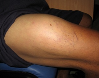
- Typically present with a large, deep, immobile, painless mass
- Thigh is the most common site
- Commonly mistaken for a hematoma or lipoma
- Physical exam should include a detailed neurovascular exam
- MRI is the preferred imaging technique
- Core-needle biopsy should be performed on any mass that is enlarging, symptomatic, deep, or > 5 cm in size
- FNA is unable to distinguish between the different sarcoma histologies
- Incisional biopsies should be orientated in the longitudinal direction to facilitate subsequent
wide local excision
- Undifferentiated pleiomorphic sarcoma is the most common subtype
- Metastatic workup consists of a chest CT
- Lymph node metastases are very rare
- Treatment of Primary Disease
- Surgery
- Function-sparing and limb-sparing operations are standard
- No role for complete compartment or muscle group resection
- Tumor should be excised with a 1 to 2 cm margin of normal tissue, without violating or rupturing
the pseudocapsule
- Previous incisional biopsy site should be excised en bloc with the specimen
- Tumors close to the skin should have an adequate skin margin excised
- Tumors that abut bone, but do not invade bone, should include periosteum in the margin
- If sacrifice of major vascular and neural structures is oncologically necessary (radical resection),
vascular and nerve reconstruction should be considered
- Radiation
- Postoperative radiation combined with limb-sparing surgery gives overall control rates equal to amputation
- Reduces risk of local recurrence from 30% to < 10%, but does not impact metastasis or overall survival
- Delivered preoperatively or postoperatively
- Postoperative radiation can be delivered as external beam or brachytherapy
- Brachytherapy allows additional radiation to a close margin (neurovascular structures) with minimal treatment
to surrounding tissues
- Preoperative radiation is associated with more wound complications, but has a better functional outcome
- Small, low-grade tumors with an adequate resection margin do not need radiation
- Chemotherapy
- Patients with large, deep, high-grade tumors may be considered for neoadjuvant or adjuvant chemotherapy
- Active agents include adriamycin and ifosfamide
- No evidence that chemotherapy improves overall survival
- Treatment of Recurrent Disease
- Local Recurrence
- Patients with isolated local recurrences should undergo re-resection
- 20% - 30% will require amputation
- Adjuvant radiation should be administered if possible
- Long-term survival is possible in two-thirds of patients
- Lung Metastases
- Most common site of metastasis
- Curative metastasectomy is possible if:
- Primary tumor is controlled
- No extrapulmonary disease
- Patient can tolerate a thoracotomy
- All lung disease is resectable
- Patients with resected pulmonary metastases have a 20% - 30% 3-year survival rate
Retroperitoneal Sarcomas
- Pathology
- Most common histologies are liposarcoma and leiomyosarcoma
- Lymphoma and metastatic testicular cancer should be considered in the differential
diagnosis of a large retroperitoneal mass
- Clinical Presentation
- Often present with nonspecific abdominal complaints: early satiety, vague discomfort,
weight loss, back or flank pain
- Physical exam may be normal or may reveal a large flank or abdominal mass
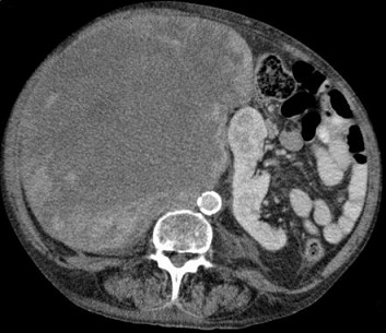
- Median size at diagnosis is 15 cm
- Diagnosis
- CT scan confirms the presence of the mass
- CT-guided biopsy is indicated if lymphoma is part of the differential diagnosis, or if
neoadjuvant therapy is being considered
- Management
- Criteria for Unresectability
- Vascular involvement of the aorta, vena cava, SMA, or iliac vessels
- Peritoneal implants
- Distant metastases that are not resectable for cure
- Involvement of the root of the mesentery and the mesenteric vessels
- Spinal cord involvement
- Surgery
- Microscopically negative (R0) resection is the goal,
but is rarely feasible due to large tumor size and anatomic constraints of the retroperitoneum
- Most resections are microscopically positive (R1)
- Kidney, colon, pancreatic tail, spleen, and psoas muscle often must be resected to achieve a
complete resection (R0/R1)
- No survival benefit to a debulking procedure (R2)
- Radiation
- Post op radiation does not reduce local recurrence
- Usually impossible to treat the entire tumor bed because surrounding structures do not tolerate
high-dose radiation well
- Neoadjuvant XRT is used in some centers for large high-grade or locally advanced tumors to optimize
local control
- Chemotherapy
- No proven benefit
- Patients with high-grade lesions may be considered for treatment
- Outcomes
- Most important prognostic factor is complete resection (R0/R1), followed by tumor grade and histologic subtype
- Majority of recurrences are local
- Most common sites of distant metastases are the liver and lungs
Desmoid Tumors
- Etiology
- Locally aggressive tumors with no potential for metastasis or malignant degeneration
- High rate of local recurrence even after complete resection
- Death may occur from local invasion of vital blood vessels or organs
- Most occur sporadically, but there are associations with familial adenomatous polyposis, pregnancy, and trauma
- Familial Adenomatous Polyposis (FAP)
- desmoid tumors occur in 10% - 20% of patients with FAP (Gardner's syndrome)
- ~50% are intraabdominal and ~ 50% involve the abdominal wall
- In FAP, complications from desmoids are the second most common cause of death (after colon cancer)
- Intraabdominal desmoids are often unresectable because they diffusely infiltrate the mesentery and
encase the mesenteric vessels
- Pregnancy
- Desmoid tumors are associated with high estrogen states
- Many tumors stop growing or even regress without intervention after delivery
- Trauma
- 30% of patients have a history of previous trauma or surgery at the tumor site
- Natural History
- Highly variable and unpredictable clinical course
- Most grow slowly and indolently over time
- Periods of growth arrest or spontaneous regression are common
- Rapid, progressive growth is less frequently seen
- Local recurrences are common, even after a complete resection
- Etiology
- Overactivation of the Wnt/beta-catenin signaling pathway appears to play a key role
- APC gene tightly regulates levels of beta-catenin in the cell
- Inactivation of the APC gene, or activating mutations in the beta-catenin gene, leads to increased levels
of beta-catenin
- Beta-catenin acts as a nuclear transcription factor which promotes cell proliferation and survival
- Clinical Presentation
- Occurs in one of three sites: trunk/extremity, abdominal wall, or intraabdominal (bowel and mesentery)
- In FAP patients, intraabdominal or abdominal wall desmoids predominate
- In sporadic cases, the shoulder girdle, hip and buttocks, and extremities are most common
- Extremity desmoids are usually located deep in the muscles
- Diagnosis
- Imaging
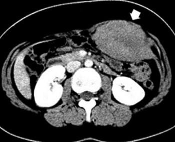
- Defines the relationship of the desmoid to adjacent structures in order to assess resectability
- MRI is preferred for trunk or extremity desmoids
- CT is preferred for intraabdominal lesions
- Imaging cannot reliably distinguish desmoids from soft tissue sarcomas
- Histologic Diagnosis
- Core biopsy is usually sufficient to make the diagnosis, although occasionally an incisional
biopsy will be required
- Desmoids lack the nuclear and cytoplasmic features of malignancy, necrosis is absent, and
mitotic figures are rare
- Treatment
- Trunk and Abdominal Wall Desmoids
- Observation
- Acceptable strategy for stable, asymptomatic lesions, especially if resection would
cause major morbidity
- Indicated in recurrent asymptomatic desmoids
- Surgery
- Indicated in symptomatic patients, or for those with progressively enlarging tumors
- Goal is complete resection with a negative microscopic margin (R0), but this is often not possible
because of anatomic constraints and the infiltrative nature of desmoids
- Skin grafts or flap reconstructions may be required for extremity desmoids
- Abdominal wall desmoids require reconstruction with mesh to minimize the risk of hernias
- Resection Margins
- Relationship between surgical margin status and local recurrence rates is unclear
- Patients with widely negative margins (R0) have recurrence rates of 16% - 39%
- Patients with microscopically positive margins (R1) have recurrence rates equivalent
to patients with R0 resections
- Patients with grossly positive margins (R2) have much higher local recurrence rates
- Radiation Therapy
- Desmoids are radiosensitive tumors
- Definitive treatment in patients who are poor surgical candidates, in patients who refuse surgery,
or in patients for whom surgical morbidity would be excessive
- Postoperative XRT
- Not recommended after an R0 resection
- Unclear whether XRT is of benefit in patients with an R1 resection
- Generally recommended after an R2 resection
- Intraabdominal Desmoids
- Surgery vs Medical Management
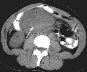
- Majority are unresectable because of diffuse involvement of the mesentery or
encasement of the mesenteric vessels
- Rate of recurrence is high after resection, as is the operative morbidity
- Multimodality medical therapy is preferred
- Medical treatments include NSAIDs (Sulindac), tamoxifen, targeted therapy (imatinib),
or chemotherapy
- Resection with small bowel transplant has been performed in several patients
Dermatofibrosarcoma Protuberans
- Etiology
- Superficial cutaneous tumor that infiltrates tissue centimeters beyond the obvious margins
- Recurs locally but rarely metastasizes
- Clinical Presentation
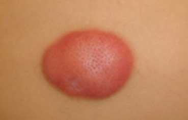
- Initially presents as an asymptomatic plaque that slowly enlarges
- Later on, the tumor becomes raised, firm and nodular
- Most common location is on the chest and shoulders
- Treatment
- Wide excision with negative margins
- May require plastic surgical reconstruction
- Radiation is not generally helpful in reducing local recurrence
References
- Sabiston, 20th ed., pgs 754 - 770
- Cameron, 13th ed., pgs 825 - 839
- UpToDate. Clinical Features, Evaluation, and Treatment of Retroperitoneal Soft Tissue Sarcoma. Mullen, John, and Delaney, Thomas. April 2018. Pgs 1 – 40.
- UpToDate. Desmoid Tumors: Epidemiology, Risk Factors, Molecular Pathogenesis, Clinical Presentation, Diagnosis, and Local Therapy. Ravi, Vinod. March 2019. Pgs 1 – 30.
- UpToDate. Dermatofibrosarcoma Protuberans. Mendenhall, William. January 2019. Pgs 1 - 28.


