Abdominal Trauma: Initial Evaluation
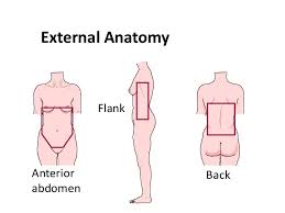
External Anatomy
- Anterior Abdomen
- defined as the area between the nipple line superiorly, inguinal ligaments and symphysis pubis
inferiorly, and the anterior axillary lines laterally
- area most assessible to physical exam and FAST exam
- Flank
- defined as the area between the anterior and posterior axillary lines from the 6th intercostal space
to the iliac crest
- Back
- defined as the area between the posterior axillary lines from the tips of the scapulae to the iliac
crests
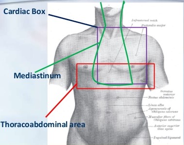
- Thoraco-abdominal
- defined as the area inferior to the nipple line anteriorly or the tips of the scapulae posteriorly,
and superior to the costal margins
- Retroperitoneum
- flank and back contain the retroperitoneal organs
- area posterior to the peritoneal lining of the abdomen
- contains the aorta, vena cava, 3rd and 4th portions of the duodenum, pancreas, kidneys and ureters,
and posterior aspects of the ascending and descending colons
- injuries to the retroperitoneum are difficult to recognize by physical exam because peritonitis may
not be present
- FAST exam or DPL do not assess this area
- Pelvis
- area surrounded by the pelvic bones, containing the lower part of the peritoneal and retroperitoneal
spaces
- contains the rectum, bladder, iliac vessels, and female reproductive organs
Mechanism of Injury
- Blunt Trauma
- spleen (40% to 55%), liver (35% to 45%), and small bowel (5% to 10%) are the most frequently injured
organs
- MVAs account for 75% of blunt abdominal injuries
- Compression Injury
- caused by a direct blow (e.g. steering wheel)
- increased intraabdominal pressure causes rupture of solid and hollow organs
- intraabdominal organs may also be crushed between the anterior abdominal wall and vertebral
column
- Deceleration Injury
- caused by falls, head-on MVAs
- results in differential movement (shearing) of fixed and movable parts of the body
- lacerations of the liver and spleen (movable) frequently occur at sites of supporting
ligaments (fixed)
- Penetrating Trauma
- Stab Wounds
- cause tissue damage by laceration or cutting
- liver (40%), small bowel (30%), diaphragm (20%), and colon (15%) are the most frequently
injured organs
- Gunshot Wounds
- low-velocity gunshot wounds cause tissue damage by laceration and cutting
- high-velocity gunshot wounds transfer more kinetic energy to abdominal viscera, cause
temporary cavitation, and may tumble and fragment, causing further injuries
- small bowel (50%), colon (40%), liver (30%), and abdominal vasculature (25%) are the most
frequently injured organs
- Blasts
- cause injury from penetrating fragment wounds and blunt injuries from the patient being
thrown or struck
- blast overpressure may cause additional pulmonary and hollow viscus injuries
Blunt Abdominal Trauma
- History
- much of the relevant history will be obtained in the field by prehospital personnel
- pertinent information includes:
- whether the patient was the driver or a passenger
- whether the patient was restrained or unrestrained, and type of restraints used
- whether the airbag deployed
- whether a prolonged extrication was required
- speed of the vehicle
- type of impact
- damage to the vehicle
- status of other passengers, including deaths at the scene
- patient’s level of consciousness and hemodynamic status during transport
- for falls, the height of the fall is important
- preexisting medical conditions and allergies
- ABCDEs
- initial resuscitation and examination proceeds according to ATLS guidelines
- priority is given to establishing an adequate airway, correcting immediately life-threatening
injuries (tension pneumothorax, hemothorax), and obtaining 2 large-bore IVs for blood and fluid
resuscitation
- x-rays of the chest and pelvis should be obtained
- gastric and urinary catheters should be inserted if not contraindicated by physical exam
- Abdominal Evaluation
- Physical Examination
- abdomen should be examined by inspection, auscultation, percussion, and palpation
- the pelvis should be assessed for stability and the rectum, perineum, penis, and vagina must
also be examined
- the abdominal exam is considered reliable and sufficient if the patient is awake, alert,
responsive, and does not have a distracting injury
- however, the abdominal exam is unreliable in many blunt trauma patients because of head
injury, intoxication, spinal cord injury, pelvic fracture, or distracting orthopedic injury
- if the abdominal examination is reliable and negative, and the patient is hemodynamically
stable, then no further studies are necessary
- patients with peritonitis on exam need a laparotomy with no further abdominal studies
- patients with an equivocal or unreliable physical exam will require further diagnostic
testing (FAST, CT scan, DPL,)
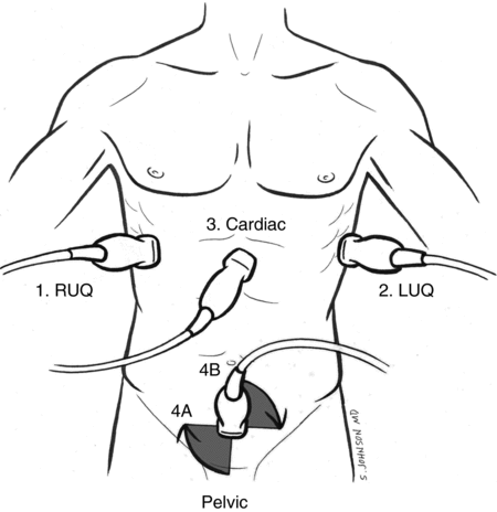
- Ultrasound (FAST)
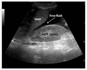
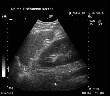
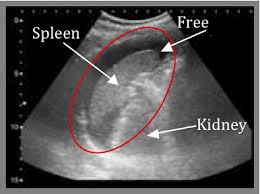
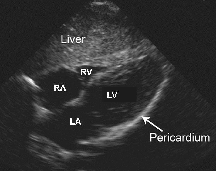
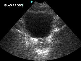
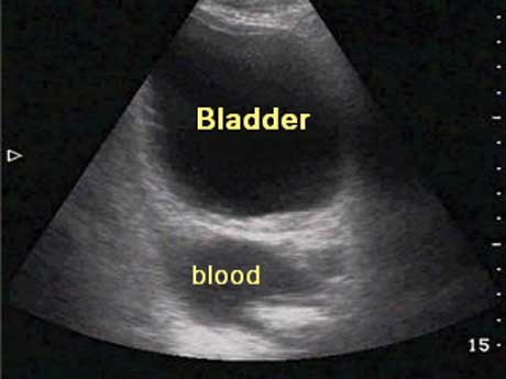
- used to detect hemoperitoneum by evaluating 4 separate areas: the pericardial sac,
hepatorenal fossa, pelvis, and the splenorenal fossa
- Advantages
- noninvasive
- rapid, inexpensive
- can be repeated frequently
- can be performed in the trauma room
- other diagnostic or therapeutic procedures may be performed simultaneously
- Disadvantages
- requires special training
- operator variability
- overlying bowel gas, obesity, or subcutaneous emphysema can make for a suboptimal
study
- a negative study does not rule out an intraabdominal injury: diaphragm, bowel, and
retroperitoneal injuries are easily missed
- a positive study is only 50% predictive of an injury that requires surgical repair
- RUQ Images
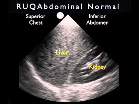
- LUQ Images
- Pericardial Images
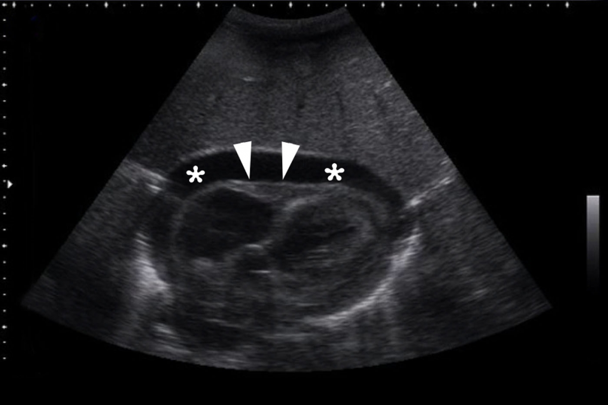
- Pelvic Images
- CT Scan
- should only be used in a hemodynamically stable patient with no obvious indication for
emergency laparotomy
- Advantages
- noninvasive
- best study to localize the site of bleeding
- can be used to grade solid organ injuries, allowing for observation of minor injuries
and decreasing the incidence of nontherapeutic laparotomies
- images the retroperitoneum, pelvis, and urinary tract well
- Disadvantages
- time-consuming; requires transport away from the ER
- poor ability to detect bowel or diaphragm injuries
- contrast allergies
- uncooperative patients
- Diagnostic Peritoneal Lavage (DPL)
- has largely been replaced by the FAST exam and CT scan
- 98% sensitive for detecting intraperitoneal bleeding
- can be performed on unstable patients in the trauma room without the need for transport
- Criteria for Positive DPL Following Blunt Trauma
- Grossly Positive
- > 10 cc blood
- enteric contents
- lavage fluid in a chest tube or bladder catheter
- Microscopically Positive
- > 100,000 RBCs
- > 500 WBCs
- amylase > serum
- bacteria, bile, vegetable matter
- Limitations of DPL
- invasive procedure with a 1% to 2% risk of iatrogenic bowel or vascular injury
(closed technique) or wound complications (open technique)
- inadequate fluid return (<250 cc) in 10%
- can not specify the exact site of bleeding
- 15% of patients undergo a laparotomy that identifies an injury that requires no
specific treatment (liver or splenic lacerations that have stopped bleeding)
- cannot evaluate the diaphragm and retroperitoneal structures
Penetrating Abdominal Trauma
- Stab Wounds
- Indications for Emergent Laparotomy
- peritonitis
- hypotension
- evisceration
- impalement
- free air or retroperitoneal air on upright chest x-ray
- blood in the rectum or stomach
- Selective Management versus Local Wound Exploration
- Selective Management
- patients without an indication for emergent laparotomy can be selectively observed,
even if the FAST exam is positive
- patients are admitted to the hospital and undergo serial exams by the same observer
for at least 24 hours
- a change in physical findings mandates further studies or surgical exploration
- Local Wound Exploration
- should only be considered if the FAST exam is negative, since a positive FAST exam
indicates peritoneal penetration
- barriers to local wound exploration include obesity, tangential wound tracts,
multiple wounds, and poor lighting and instruments in the ER
- Negative Wound Exploration
- wound does not penetrate the anterior rectus or external oblique fascia
- wound may be closed and the patient discharged from the ER
- Positive Wound Exploration
- if an exploration is positive or equivocal, then the patient should be
admitted for observation
- if the patient’s exam changes, then a decision must be made regarding
immediate laparotomy or additional diagnostic studies
- for anterior abdominal stab wounds, laparoscopy can be used to determine if
the peritoneum has been violated, since CT scan is less useful in these
cases because it misses bowel injuries
- for flank and back stab wounds, double or triple contrast CT is the study of
choice
- Thoracoabdominal Stab Wounds
- stab wounds between the nipples and costal margin
- selective management risks missing a diaphragmatic injury while local wound exploration
carries a risk of pneumothorax
- laparoscopy or DPL are potential diagnostic studies to rule out diaphragmatic injury
- Gunshot Wounds (GSWs)
- all wounds should be identified with radiopaque markers, and x-rays should be obtained to determine
their relation to missile position
- the number of skin wounds and missiles should add up to an even number
- Anterior Abdomen GSWs
- great majority of patients, even if hemodynamically stable, require exploratory laparotomy
because of the high risk of injury
- selective management in hemodynamically stable patients has been described for right upper
quadrant GSWs, and morbidly obese patients without evidence of peritoneal penetration on CT
scan
- Flank and Back GSWs
- CT scan can delineate the bullet track and diagnose associated injuries in stable patients
Exploratory Laparotomy in Trauma
- OR Setup
- OR should be large enough to accommodate more than one surgical team in case simultaneous procedures
are necessary
- rapid transfusion device and blood should be available
- room temperature should be able to go as high as 80 degrees
- patient must be prepped from the neck to mid thighs
- Operative Imperatives
- Hemorrhage Control
- first priority
- direct pressure or proximal vascular control
- all four abdominal quadrants are packed with laparotomy pads
- Control of Contamination
- obvious leakage is controlled with clamps, staples, or sutures
- Identify all Injuries
- inspect the entire bowel and intraabdominal colon
- evaluate the diaphragm
- enter the lesser sac to inspect the pancreas and posterior wall of the stomach
- Definitive Repair of Injuries
- may defer if the patient is too unstable (damage control laparotomy)
References
- ATLS, 10th ed., pgs 82 - 101
- Cameron, 11th ed., 1010 – 1012, 1021 – 1027
- Sabiston, 20th ed., pgs 432 - 435


